Normal Rv Pressure Tracing
Right And Left Heart Hemodynamics Thoracic Key

Right Heart Catheterization Pcipedia

Cv Physiology Pulmonary Capillary Wedge Pressure

Figure 2 Differentiation Of Constriction And Restriction Jacc Journal Of The American College Of Cardiology

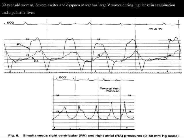
Ra Cvp Waveform Interpretation Rk Md
Right Ventricular Pressure Tracing Patient 9 Thin Arrow Download Scientific Diagram
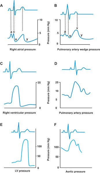
This pressure waveform looks similar to the rv except the diastolic will be a bit higher.
Normal rv pressure tracing.

Right Heart Catheterization Cheat Sheet Interpretation Of Pressures And Waveforms Direct Measurements

Cv Physiology Cardiac Cycle

Normal Jugular Venous Waveform Jugular Venous Pressure Jugular Liver Failure

Fig 5 Measurement Of Pressure Gradients In Mitral Stenosis Tracing The Continuous Wave Doppler Velocity Time I Cardiac Sonography Cardiac Cycle Mitral Valve
Source : pinterest.com
